-
-
-
-
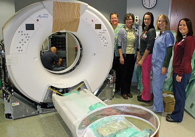 Susan Bender, Monica Stark, Stacia Johnson, Maria Davis and Jill Henkle pose near the new CT.
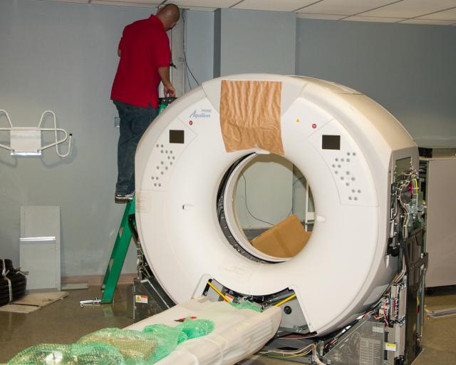
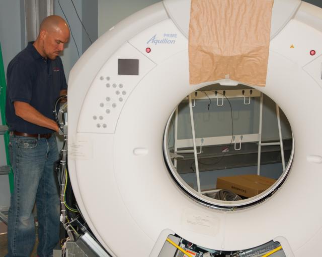
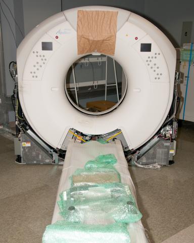
The Virginia Gay Hospital’s Imaging Services Department (X-Ray) is installing a new CT (Computerized Tomography) machine this week. For the past couple of weeks technicians have removed the former machine, leveled floors and today the new machine is being installed. In the interim, patients needing CT scans are using a mobile system parked behind the hospital.
Monica Stark, Imaging Services Coordinator, said there are tremendous benefits to the unit, for example: higher imaging speeds (e.g., the former machine imaged 32 slices/rotation compared to 40 per rotation with the new model) and that means lower radiation dose for patients; decreased reconstruction times (i.e., the time it takes for the images to be ready for the physician to view has decreased substantially from 10-15 minutes to 2-3 minutes) meaning quicker reading times; larger gantry opening (i.e., larger hole) for patients; and there are considerable cost savings with this new system.
The new machine should be operational within the next week.
What is CT?
Although also based on the variable absorption of x rays by different tissues, computed tomography (CT) imaging, also known as "CAT scanning" (Computerized Axial Tomography), provides a different form of imaging known as cross-sectional imaging. The origin of the word "tomography" is from the Greek word "tomos" meaning "slice" or "section" and "graphe" meaning "drawing." A CT imaging system produces cross-sectional images or "slices" of anatomy, like the slices in a loaf of bread. The cross-sectional images are used for a variety of diagnostic and therapeutic purposes.
How a CT system works
-A motorized table moves the patient through a circular opening in the CT imaging system.
-As the patient passes through the CT imaging system, a source of x rays rotates around the inside of the circular opening. A single rotation takes about 1 second. The x-ray source produces a narrow, fan-shaped beam of x rays used to irradiate a section of the patient's body (Figure 4). The thickness of the fan beam may be as small as 1 millimeter or as large as 10 millimeters. In typical examinations there are several phases; each made up of 10 to 50 rotations of the x-ray tube around the patient in coordination with the table moving through the circular opening. The patient may receive an injection of a "contrast material" to facilitate visualization of vascular structure.
-Detectors on the exit side of the patient record the x rays exiting the section of the patient's body being irradiated as an x-ray "snapshot" at one position (angle) of the source of x rays. Many different "snapshots" (angles) are collected during one complete rotation.
-The data are sent to a computer to reconstruct all of the individual "snapshots" into a cross-sectional image (slice) of the internal organs and tissues for each complete rotation of the source of x rays.
Source: FDA


Comments
Submit a CommentPlease refresh the page to leave Comment.
Still seeing this message? Press Ctrl + F5 to do a "Hard Refresh".